Details
The Science of X-ray Imaging: Why It Works
Introduction
X-ray imaging is arguably the most advanced medical and industrial science with non-surgical testing of internal shapes. From the diagnosis of fracture to detection of hidden structural defects in industrial products, X-ray technology has been the unbridled master power of contemporary science. What is X-ray imaging all about? Take a look at the science of X-ray imaging, the manner in which it is carried out, the whys of it being carried out, and what is new with X-ray technology.
What Are X-rays?
X-rays are a form of short-wavelength, high-energy, electromagnetic radiation, which come in between ultraviolet light and gamma rays in the category of electromagnetic radiations. X-rays have evil powers of penetration and can pass even through the bulk of materials quite easily, because of which they rule very enormously in the imaging industry.
Discovery of X-rays
Wilhelm Conrad Roentgen was a German physicist who in 1895 was attempting to conduct an experiment of observing cathode rays and in the process stumbled upon X-rays. He understood that the rays would be capable of passing through solids and producing dark shadows on photographic plates. He was the first to receive the Nobel Prize in Physics in 1901 and the creator of modern radiographic imaging.
How X-ray Imaging Works
The X-ray imaging procedure consists of three basic components:
1. X-ray Source – Where X-rays are made.
2. Object or Body Part – To be X-rayed.
3. X-ray Detector – Film or digital detector to capture transmitted X-rays.
1. X-ray Generation
X-rays are produced in an X-ray tube, a vacuum glass tube with a cathode (negative electrode) and an anode (positive electrode). The major steps involved in X-ray generation are:
- Electron Emission: Electron emission from the cathode at high voltage.
- Acceleration: Acceleration of electrons to high speed in the anode direction.
- X-ray Generation: X-rays produced due to collision of high-speed electrons on the anode by two basic processes:
- Bremsstrahlung Radiation: As a result of electrons deceleration when colliding with the target.
- Characteristic Radiation: As a result of electrons colliding with atoms and inner-shell electron transitions. It is a static electricity effect.
2. Interaction with the Object
When X-rays pass through an object (human body or material to be X-rayed by industry, etc.), the tissue will scatter X-rays as follows:
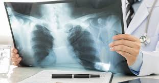
- Bony hard tissue will refract X-rays and will be white on an X-ray film.
- Organic soft tissue like muscles will absorb less energy from X-rays and will be gray.
- Air cavities (the lungs, for example) permit most X-rays to travel through and therefore are black.
This differential absorption contrast creates contrast to X-ray images, and therefore one can see inside.
3. Recording the X-ray Image
Once the X-rays have traveled through the object, they are recorded using X-ray film (analog) or detectors (digital radiography, DR). The stored X-ray image is processed for display.
Types of X-ray Imaging Modes
1. Routine X-ray Radiography
It is the most frequent X-ray imaging method employed in the diagnosis of fracture, infection, and tumor.
2. Computed Tomography (CT Scan)
CT scan takes a series of X-ray images at different angles to form a 3D cross-section image of the body for the purpose of deep imaging of bones, organs, and tissues.
3. Fluoroscopy
A real-time video imaging of X-rays method applied in the angiography procedure, barium swallow tests, and catheter placements.
4. Mammography
An X-ray imaging method applied for early breast cancer detection.
5. Industrial X-ray Imaging
Industrial X-ray imaging is applied for non-destructive testing (NDT) and allows for weld inspection, structural flaw inspection, and composite material inspection.
Advantages of X-ray Imaging
1. Non-Surgical Diagnosis – Complete details without surgery with X-rays.
2. Easy and Fast – Conveniently and comfortably available X-ray imaging.
3. Affordability – Less costly than MRI and CT scans.
4. Multifaceted – Both business and medical applications.
Recent Development in X-ray Technology
1. Digital Radiography (DR)
Unlike the old X-ray film-based imaging, Digital Radiography (DR) uses advanced flat-panel detectors to produce images instantly, with improved resolution, and lower radiation.
2. Portable X-ray Machines
Such devices like MIN-X Handheld X-ray Systems enable bedside diagnosis, emergency medical imaging, and use in remote areas.
3. Artificial Intelligence in X-ray Analysis
Computer software nowadays assist radiologists to identify abnormalities, fractures, and early sign of disease accurately and rapidly.
4. Low-Dose X-ray Imaging
Future technology advances will offer smaller radiation dose with improved imaging resolution, offering diagnostic and safe treatment.
5. 3D X-ray Imaging
New third-dimensional radiographic imaging devices offer hierarchy of structural analysis by depth, and orthopedics, dentistry, and industrial inspection are merely a few instances of applications where they can be used.
Safety Precautions for X-ray Imaging
Even though X-ray imaging is a required diagnostic process, safety measures must be adhered to in order to deliver the least possible radiation exposure:
1. Lead Shielding – Sensitive organs shielded from unnecessary exposure.
2. Minimum Exposure Time Limitation – Limiting X-ray scanning to the absolute minimum required.
3. Maximum Equipment Calibration – Routine equipment calibration to peak efficiency and proper output.
4. ALARA Principle – Maintain the level of radiation As Low As Reasonably Achievable.
Conclusion
X-ray imaging technology has transformed medical diagnosis and industrial inspection to provide non-invasive high-precision tests. From the detection of fractures to aerospace part inspection, X-ray technology is evolving with digital radiography, AI uptake, and 3D imaging. With safety, efficiency, and technological improvement, X-ray imaging fuels modern diagnostics and quality control.
We at Star Nuke are committed to innovative X-ray solutions, where enhanced diagnostic accuracy is supported by innovative portable X-ray machines and digital radiography. Contact us today and find out how technology can transform your imaging needs.

