
Details
The Early Days of X-Ray Technology
The initial X-ray photograph—Roentgen's wife's hand—initiated a new age of medical imaging, an era that would revolutionize the process. Imaging in the human body without the necessity of surgery introduced new options in diagnosis and in planning treatments. Early X-ray machines were rough and risky when it came to radiation, for little was yet known about the protection from radiation. Protective clothing was largely not available, patients and physicians were subjected to full hazards.
Despite all these challenges, X-ray technology later prospered in medical diagnosis. In the early 20th century, X-rays were used in war medicine to detect bullets and fractures, which significantly improved war medical treatment. The invention of contrast agents further improved the use of X-rays such that doctors could see soft tissues such as blood vessels and the gastrointestinal tract.
Advancements in Medical Imaging
The contemporary X-ray technology has developed far more than the earlier one with increased safety, efficiency, and image quality. Digital radiography (DR) and computed radiography (CR) have replaced ancient film-based technology with real-time feedback imaging using decreased doses of radiation. Developments such as fluoroscopy and CT scans have also expanded the range of medical uses of X-rays with the capability to provide real-time and cross-sectional images of the body with high accuracy.
CT scans, which emerged in the 1970s, revolutionized diagnostic imaging since they allowed doctors to view clear, three-dimensional images of internal structure. The innovation significantly increased the precision in diagnosing diseases such as tumors, internal hemorrhage, and complex fractures. Low-dose X-ray technology has also minimized the exposure to radiation, making imaging patient-safe, particularly for pediatric and follow-up imaging purposes.
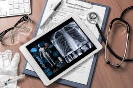
Furthermore, the use of artificial intelligence (AI) in medical imaging has improved diagnostic performance. AI software is able to scan X-ray images to identify abnormalities like fractures, tumors, and lung disease with unmatched accuracy. This technology is especially useful in rural or underserved areas, where specialty radiologists are not readily available.
Industrial and Security Applications
Outside the field of medicine, X-ray technology has important industrial and security uses. In non-destructive testing (NDT), X-ray inspection guarantees the integrity of essential structures from pipelines to aircraft components. Preventing catastrophic structural failure and collapse through internal flaws detection, i.e., voids or cracks, X-ray-based NDT increases reliability and safety in various industries.
X-ray imaging is of paramount importance in security to identify threats. X-ray imaging equipment placed in airport screening machines, border protection, and government agencies utilize X-ray imaging to scan for weapons, explosives, and prohibited drugs to attain public security. Next-generation dual-energy X-ray scanners are able to distinguish between organic and inorganic substances and aid security personnel in identifying suspected threats more effectively.
X-ray fluorescence technology is one more significant application, used in forensic science, archaeology, and material analysis. XRF helps scientists identify the elemental composition of items, providing informative data in uses ranging from criminal investigation to verifying historical artifacts.
X-Ray in Research and Space Exploration
X-ray imaging does not stop in medicine and industry; it is also a vital tool in scientific investigation and space travel. X-ray telescopes, such as the NASA Chandra X-ray Observatory, allow astronomers to study high-energy cosmic phenomena, including black holes, neutron stars, and supernovae. These studies have provided us with the most valuable information about the universe's most violent environments, and they have taught us more about cosmic evolution and astrophysical processes.
In materials science, X-ray diffraction (XRD) assists scientists in the study of the atomic material structure, driving nanotechnology innovation, medicine, and electronics. XRD is also the central part of medicine discovery because it assists researchers in learning about the crystal structure of new drugs, improving drug design and performance.
The Future of X-Ray Technology
A new generation of technologies is on the horizon that are taking X-ray imaging to a new level. Artificially intelligent image analysis, X-ray machines in portable packages, and spectral imaging techniques will transform diagnosis and industrial inspection. Portable X-ray machines, for instance, are transforming emergency medicine by enabling quick on-site imaging in disaster zones, remote locations, and even in the field.
Photon-counting CT scanners are another development, with improved image resolution from the use of lower doses of radiation. They can differentiate between various tissue types to an extent not previously possible, with more detection of cancer and osteoporosis at an early stage. Spectral imaging, where an array of levels of X-ray energy are detected, can provide greater accuracy of material differentiation both medically and industrially.
In addition, phase-contrast X-ray imaging technology can potentially transform medical diagnosis. Traditional X-ray imaging exploits tissue absorption differences, while phase-contrast imaging can detect small variations in refractive index and produce high-contrast images of soft tissues. The method can potentially extend disease detection at an early stage by a significant degree, e.g., lung cancer and breast cancer.
Sustainability is also being addressed in the design of X-ray technology. Steps are being made to design environmentally friendly X-ray systems that generate less electronic waste and have lower levels of energy consumption. New lead-free materials are being explored to offer more effective radiation protection with less environmental degradation.